


In order to determine the type of stroke being suffered, a general physical examination is necessary to find out what the symptoms are or were, when they started, and what the patient was doing when the symptoms began.
The physician will ask if any medications are currently being taken, and a personal and family history of heart disease, transient ischemic attack, or stroke will be recorded. It is also important to know whether the patient has experienced any recent head injuries.
Blood pressure will be checked, along with the heart and carotid arteries, which may indicate atherosclerosis, also known as cardiovascular disease. The physician may also use an ophthalmoscope (a familiar device used in eye exams and regular checkups) to check for signs of tiny cholesterol crystals or clots in the blood vessels at the back of the eyes.
Emergency treatment for stroke depends on whether you’re having an ischemic stroke, a hemorrhagic stroke, or Transient Ischemic Stroke (TIA). Once the initial exam is complete, specialized tests may be needed to determine the affected area of the brain and/or location of a clot.
These tests may include:
Blood Tests / Lab Work
Blood Tests will tell the medical team how fast the blood is clotting, whether the blood sugar is abnormally high or low, whether critical blood chemicals are out of balance, and whether there is an infection. Managing the blood’s clotting time and levels of sugar and other key chemicals will also be part of stroke aftercare.
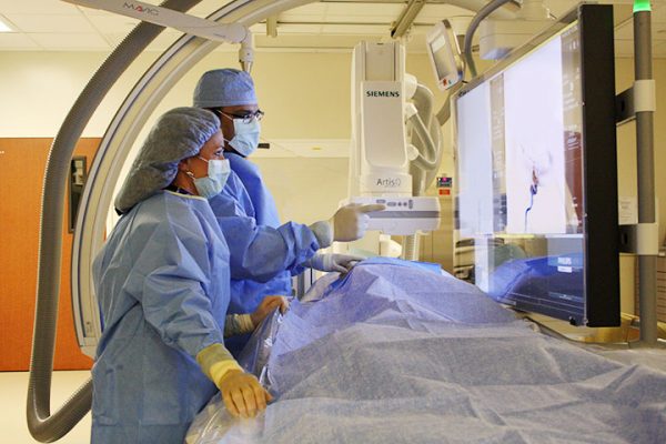
Cerebral Angiogram with Digital Subtraction Angiography (DSA)
Cerebral Angiogram with Digital Subtraction Angiography (DSA) uses complex, computerized x-ray technology to produce three-dimensional-like images of the arteries. DSA is a technique of imaging that can allow the doctor to view real-time moving images of blood vessels beneath the bony skull.
CT Scan/CT Perfusion
A computerized tomography (CT) scan uses a series of X-rays to create a detailed image of the brain. It can show a hemorrhage, tumor, a stroke, or other conditions. A computerized tomography angiography uses injected dye into the bloodstream to view the blood vessels in the neck and brain in greater detail.
CTA Computed Tomography Angiography
Computed tomography angiography (CTA) is similar to a CT scan and uses an IV contrast medium which allows many different x-rays to be taken at different angles. This allows rapid evaluation of the extent of the stroke and the amount of brain tissue involved.
Cartoid Doppler Ultrasounds
In a carotid doppler ultrasound, the arteries in the neck (the carotid arteries) that supply blood to the brain are visualized using high-frequency sound waves. These detailed images are used to find evidence of plaque build-up in the arteries, but is limited in what it can view.
Cerebral Angiogram
In a cerebral angiogram, a catheter is inserted through a small incision, usually in the groin, and guided through the major arteries into the carotid or vertebral artery. The doctor then injects a dye into the blood vessels to make them visible under X-ray imaging. This procedure gives a detailed view of arteries in the brain and neck.
ECHOCardiogram
An echocardiogram uses sound waves to create detailed images of the heart. It can find a source of clots in the heart that may have traveled from the heart to the brain and caused the stroke. In a transesophageal echocardiogram, the doctor inserts a catheter, with a small transducer attached, into the throat and down into the esophagus to the stomach. Because the esophagus is directly behind the heart, this procedure can create clear, detailed ultrasound images of the heart and any blood clots.
Lumbar Puncture
A lumbar puncture, also known as a spinal tap, is a procedure where a needle is used to collect cerebrospinal fluid from within the spine. Diagnostic tests of the fluid can indicate if there is bleeding in or around the brain.
Magnetic Resonance Imaging (MRI) and Magnetic Resonance Angiogram (MRA)
Magnetic Resonance Imaging (MRI) and Magnetic Resonance Angiogram (MRA) use powerful radio waves and magnets to create a detailed view of the brain. An MRI can detect brain tissue damaged by an ischemic stroke and brain hemorrhages. In magnetic resonance angiography and magnetic resonance venography, dye is injected into a blood vessel to view the arteries and veins and highlight blood flow.
Transcranial Doppler (TCD)
In Transcranial Doppler (TCD), ultrasound waves are transmitted through the tissues of the skull. These sound waves reflect off of the blood cells moving within the blood vessels, allowing the radiologist to calculate their speed.
Identifying the type of stroke will determine the type of treatment that will best serve the patient.